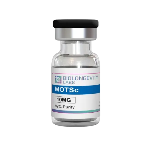
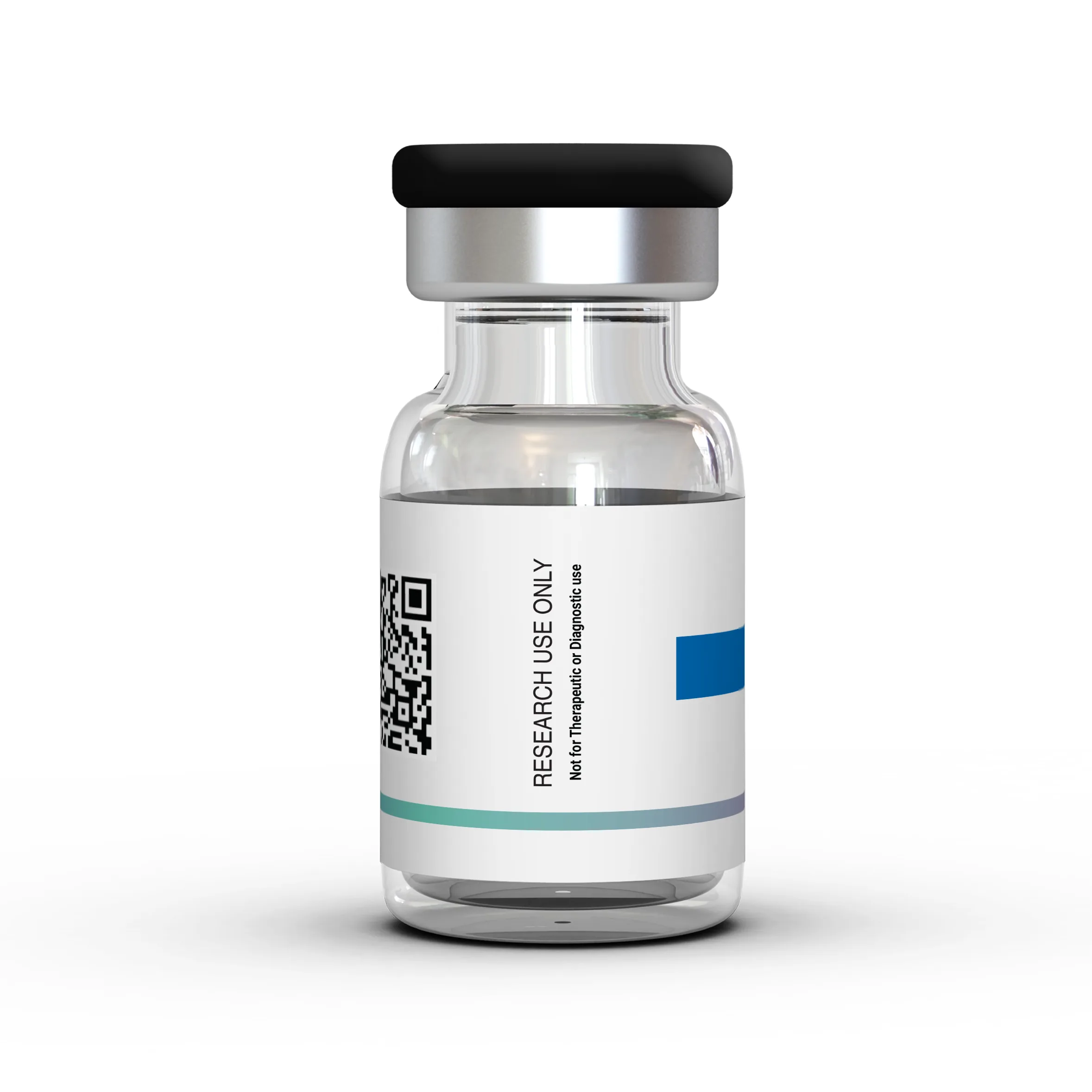
MOTS-c 10mg is a synthetic 16-residue peptide whose sequence matches a short open reading frame inside the mitochondrial 12S rRNA gene. It is made by controlled solid-phase peptide synthesis and supplied as a lyophilized powder in a sealed 10mg vial. Each vial is third-party tested by HPLC to ≥99% purity, with a Certificate of Analysis available. Laboratories use MOTS-c to study AMPK signaling, folate and purine metabolism, and how a mitochondrially encoded peptide can relay signals to the nucleus.
≥99% Purity
Third-Party Tested
COA Available
U.S. Manufactured
Quantity
In stock
Third-Party Verified Quality
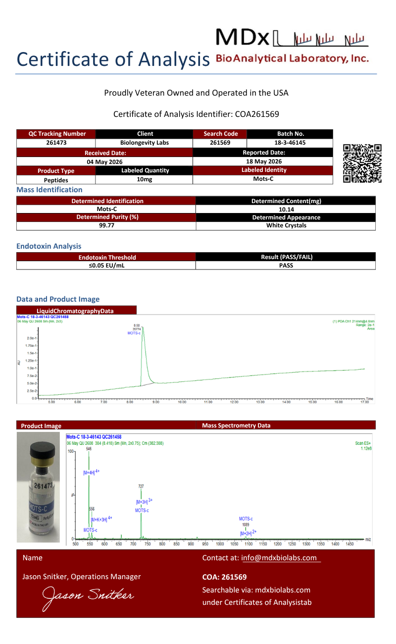
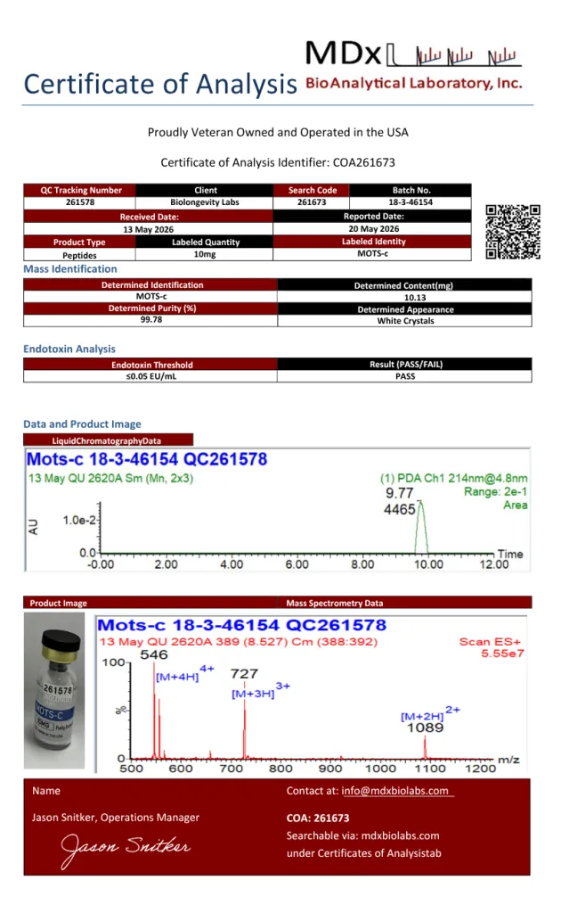
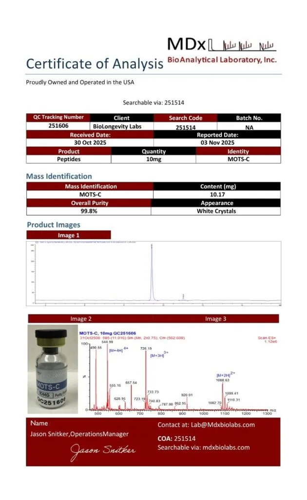
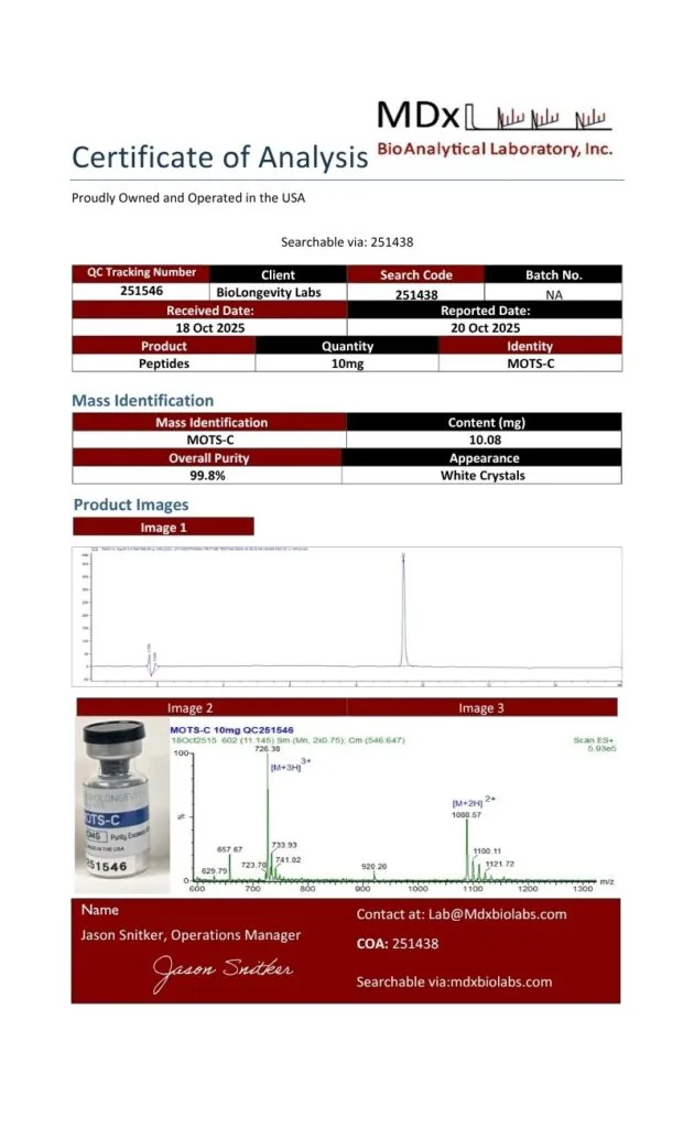
≥99% purity verified through independent UHPLC-MS testing. Certificates of Analysis are available before purchase.
COAs available before purchase
U.S.-manufactured research-grade
Same-business-day U.S. dispatch for in-stock orders placed before 12 PM PT
Secure Checkout · Free shipping on orders over $400
MOTS-c stands for “mitochondrial open reading frame of the 12S rRNA type-c.” It was first described in 2015 as a member of the small family of mitochondrial-derived peptides, signaling molecules encoded within mitochondrial DNA [1].
Most cellular proteins are encoded in nuclear DNA. MOTS-c is unusual because its code sits in the mitochondrial genome, which makes the MOTS-c peptide a useful tool for questions about how mitochondria and the nucleus communicate.
BioLongevity Labs supplies MOTS-c as the free-acid, linear form of the sequence MRWQEMGYIFYPRKLR, with no disulfide bridges. The vial holds the parent peptide only, with no fillers or carrier blends.
MOTS-c, MOTSc, Mitochondria-derived peptide MOTS-c, mitochondrial open reading frame of the 12S rRNA-c
Peptide Class
Mitochondrial-derived peptide (MDP)
Source
Synthetic (solid-phase peptide synthesis)
Vial Content
10mg
Purity
≥99% (HPLC)
Appearance
White lyophilized powder
Solubility
Soluble in bacteriostatic water
Storage
-20°C, sealed, protected from light and moisture
Storage and Handling
Store the lyophilized compound at -20°C in its sealed vial, away from light and moisture.
After reconstitution, store at 2–8°C and use promptly.
Avoid repeated freeze-thaw cycles. Aliquot reconstituted material if it will be used across several experiments.
Maintain aseptic handling to preserve compound integrity.
Lyophilized Format
This compound ships in lyophilized (freeze-dried) form. Freeze-drying supports long-term storage stability and preserves compound integrity. No fillers are added.
Research Use Disclaimer
MOTS-c is supplied for research use only. It is not a drug, food, cosmetic, or dietary supplement and has not been evaluated by the FDA. By purchasing, the buyer confirms the compound will be used solely for in vitro research.
Frequently Asked Questions
What Is MOTS-c 10mg?
MOTS-c 10mg is a vial containing 10 milligrams of synthetic MOTS-c, a 16-residue mitochondrial-derived peptide supplied as a lyophilized powder. BioLongevity Labs sells it for in vitro research on AMPK signaling and mitochondria-to-nucleus communication.
What Is the Amino Acid Sequence of MOTS-c?
The MOTS-c sequence is MRWQEMGYIFYPRKLR, or Met-Arg-Trp-Gln-Glu-Met-Gly-Tyr-Ile-Phe-Tyr-Pro-Arg-Lys-Leu-Arg. It is a linear 16-residue peptide with a free C-terminal acid and no disulfide bridges.
What Are the CAS Number and Molecular Weight of MOTS-c?
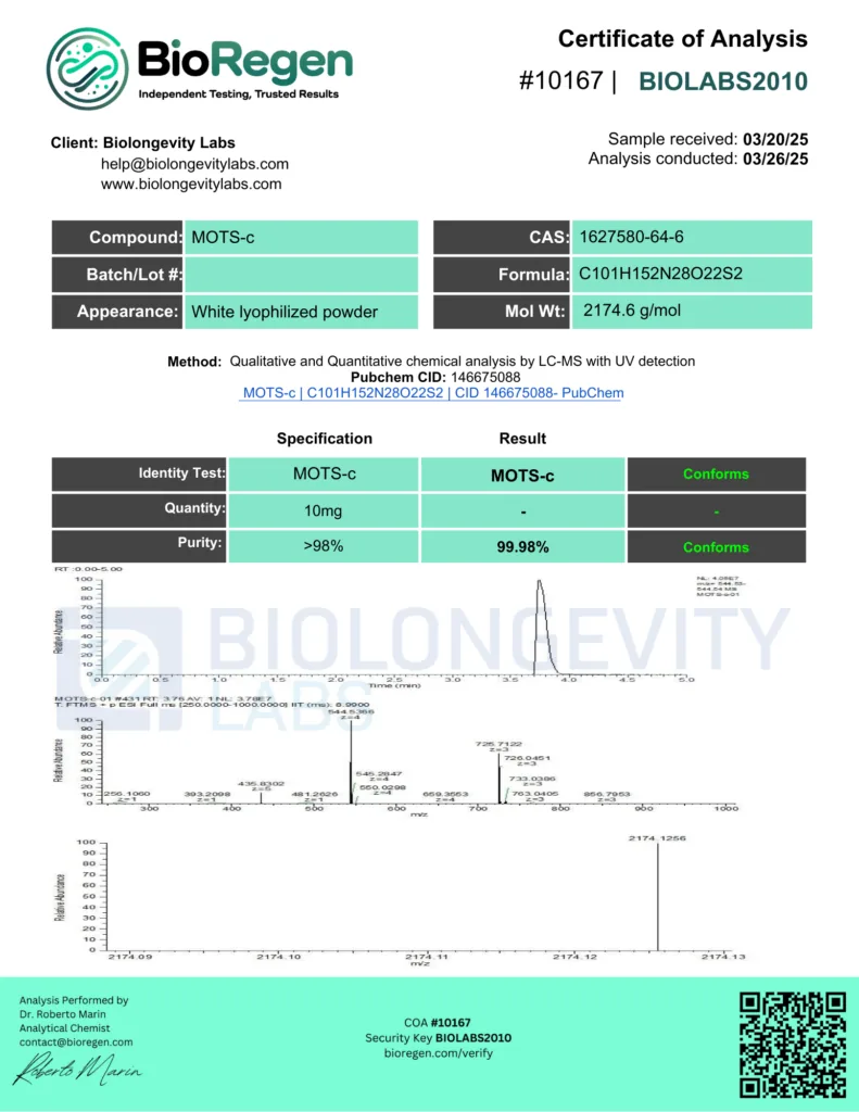
MOTS-c is registered under CAS number 1627580-64-6 and PubChem CID 146675088. Its molecular formula is C₁₀₁H₁₅₂N₂₈O₂₂S₂, with a molecular weight of 2174.6 g/mol.
How Should MOTS-c Be Stored?
Lyophilized MOTS-c should be kept sealed at -20°C, protected from light and moisture. Once reconstituted, it should be held at 2–8°C, used promptly, and kept away from repeated freeze-thaw cycles.
How Is the Purity of MOTS-c Verified?
MOTS-c from BioLongevity Labs is third-party tested by HPLC to ≥99% purity. A Certificate of Analysis is available for the product.
Research Overview
MOTS-c was identified in 2015 when a short open reading frame within the mitochondrial 12S rRNA gene was found to encode a 16-amino-acid peptide [1]. In cultured cell lines, the peptide inhibited the folate cycle and the de novo purine synthesis tied to it. That shift led to activation of AMPK, the cell’s central energy sensor [1]. In mouse studies, skeletal muscle appeared to be its primary target tissue [1].
A 2018 study added a second layer. Under glucose restriction and oxidative stress, MOTS-c moved from the cytoplasm into the nucleus in an AMPK-dependent manner [2]. There it regulated a broad set of genes, including those carrying antioxidant response elements, and interacted with the stress-responsive transcription factor NRF2 [2]. A later review describes this as direct mitochondrial-encoded control of nuclear gene expression, a reversal of the usual nucleus-to-mitochondria direction [3].
The research threads below show how laboratories have extended these findings in muscle-cell and sequence-level models.
Muscle-Cell and AMPK Pathway Research
In C2C12 myotubes, blocking AMPK with Compound C lowered both PGC-1α and MOTS-c expression, while PGC-1α knockdown reduced MOTS-c levels [4]. Adding recombinant MOTS-c raised MOTS-c expression in the same cells, which points to an AMPK and PGC-1α feedback loop [4]. Separate work in mouse models of different ages linked MOTS-c to nuclear genes for metabolism and proteostasis, and to myoblast adaptation under metabolic stress [5].
Sequence Variant Research
A naturally occurring variant, K14Q, replaces lysine at position 14 with glutamine. Researchers have described it as a form of MOTS-c with reduced biological activity, which makes it a useful comparison point for structure and activity studies [6].
Research Area
In Vitro Application
AMPK Signaling
Phospho-AMPK readouts in cultured myotubes and other cell lines exposed to MOTS-c
Folate and Purine Metabolism
Metabolite profiling of folate-cycle and de novo purine pathway intermediates
Mitochondria-to-Nucleus Signaling
Imaging of MOTS-c nuclear translocation under glucose restriction or oxidative stress
Stress-Response Gene Expression
NRF2 interaction and antioxidant response element gene panels
Myoblast Metabolic Stress
Adaptation assays in C2C12 myoblasts and myotubes
Structure and Activity
Side-by-side comparison of the parent sequence with the K14Q variant
References
Lee C, Zeng J, Drew BG, et al. (2015). The mitochondrial-derived peptide MOTS-c promotes metabolic homeostasis and reduces obesity and insulin resistance. Cell Metabolism, 21(3), 443–454. https://pmc.ncbi.nlm.nih.gov/articles/PMC4350682/
Kim KH, Son JM, Benayoun BA, Lee C. (2018). The Mitochondrial-Encoded Peptide MOTS-c Translocates to the Nucleus to Regulate Nuclear Gene Expression in Response to Metabolic Stress. Cell Metabolism, 28(3), 516–524.e7. https://pmc.ncbi.nlm.nih.gov/articles/PMC6185997/
Yang B, Yu Q, Chang B, et al. (2021). MOTS-c interacts synergistically with exercise intervention to regulate PGC-1α expression, attenuate insulin resistance and enhance glucose metabolism in mice via AMPK signaling pathway. Biochimica et Biophysica Acta. Molecular Basis of Disease, 1867(6), 166126. https://doi.org/10.1016/j.bbadis.2021.166126
Reynolds JC, Lai RW, Woodhead JST, et al. (2021). MOTS-c is an exercise-induced mitochondrial-encoded regulator of age-dependent physical decline and muscle homeostasis. Nature Communications, 12(1), 470. https://pmc.ncbi.nlm.nih.gov/articles/PMC7817689/
Kumagai H, Natsume T, Kim SJ, et al. (2022). The MOTS-c K14Q polymorphism in the mtDNA is associated with muscle fiber composition and muscular performance. Biochimica et Biophysica Acta. General Subjects, 1866(2), 130048. https://pmc.ncbi.nlm.nih.gov/articles/PMC8741734/
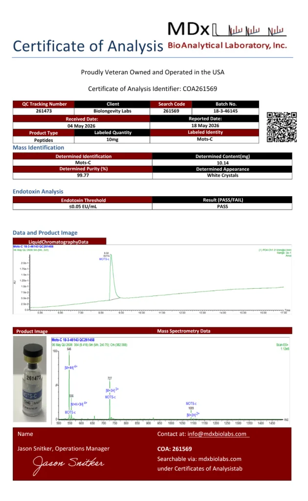
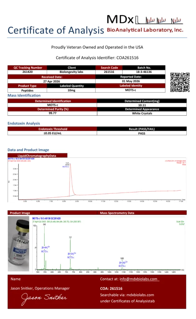
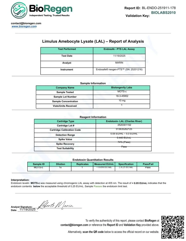
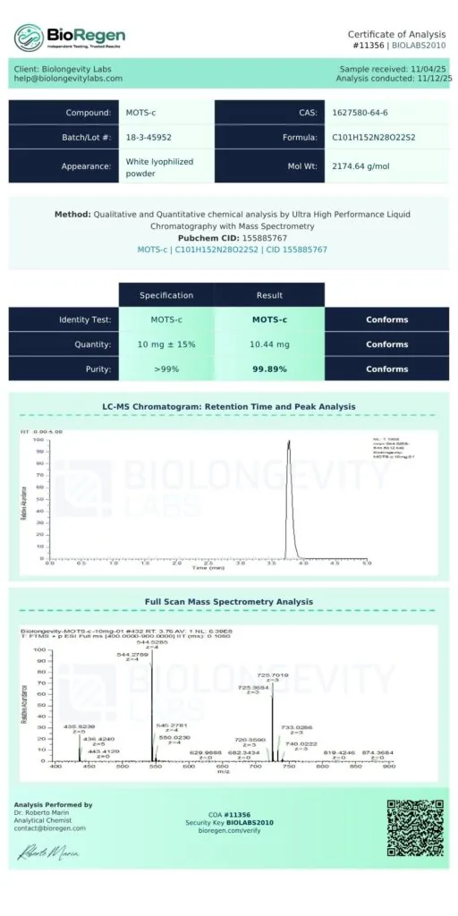
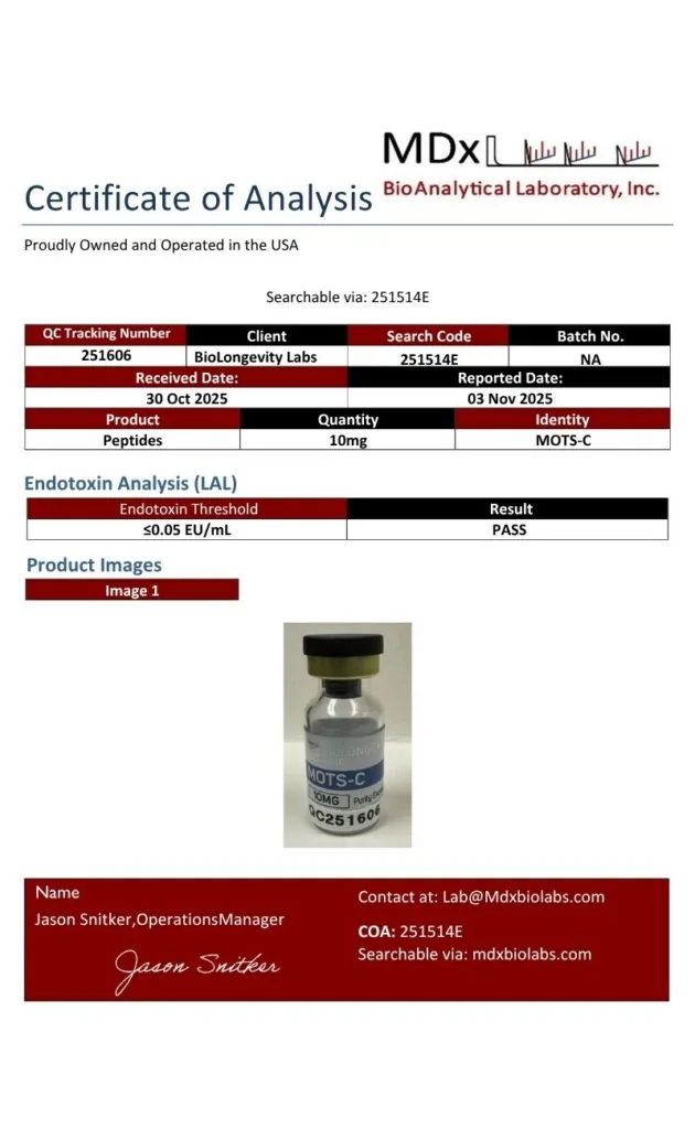
A Certificate of Analysis (COA) is a document that verifies a compound’s identity, purity and quality through independent laboratory testing. Every compound from BioLongevity Labs is third-party tested, and its COA is published here, so researchers can review the testing record before a compound enters a protocol.
Each COA reports results from third-party laboratory analysis, including:
Ultra-high-performance liquid chromatography with mass spectrometry (UHPLC-MS) for purity, measured against a 98% or 99% specification depending on the compound
Mass identification for molecular confirmation and content quantitation
Endotoxin testing by Limulus amebocyte lysate (LAL) or recombinant Factor C (rFC) assay, depending on the laboratory
Visual and physical characterization of the finished material
How to Verify a COA Independently
You can check every certificate against the issuing laboratory’s own records, not just the copy hosted here. Verification does not depend on BioLongevity Labs.
AV Analytics certificates carry a search code. Scan the QR code on the certificate to open the laboratory’s verification page, or enter the search code at avanalytical.com/coa-search to pull the official record.
BioRegen reports of analysis carry a Report ID and a Validation Key. Scan the QR code on the certificate to open the official record, or quote both identifiers when you contact the laboratory at the address printed on the report.
SafeCert Labs issued a number of earlier certificates. Each one carries a COA number and the signature of the reporting chemist. Quote that number when you ask the laboratory to confirm it.
Older certificates from other laboratories list their own verification details on the document.
When a batch is sent to more than one laboratory, each certificate resolves at its own issuing laboratory. A researcher can then check the results against two unrelated sources.
These COAs come from independent certified laboratories, not in-house testing. That gives researchers a verifiable record of molecular integrity for each compound. All compounds are supplied for research use only.
Scientifically reviewed by Dr. Ky H. Le, MD. Dr. Le is a board-certified family medicine physician with over 20 years of clinical experience. Dr. Le validates the scientific accuracy of all technical content and research citations.
Disclaimer: For Research Purposes Only
This content is provided strictly for research purposes and does not constitute an endorsement or recommendation for the non-laboratory application or improper handling of peptides designed for research. The information, including discussions about specific peptides and their researched benefits, is presented for informational purposes only and must not be construed as health, clinical, or legal guidance, nor an encouragement for non-research use. Peptides described here are solely for use in structured scientific study by authorized individuals. We advise consulting with research experts, medical practitioners, or legal counsel prior to any decisions about obtaining or utilizing these peptides. The expectation of responsible, ethical utilization of this information for legitimate investigative and scholarly objectives is paramount. This notice is dynamic and governs all provided content on research peptides.