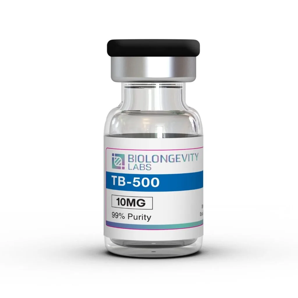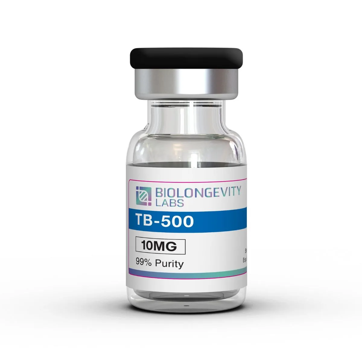
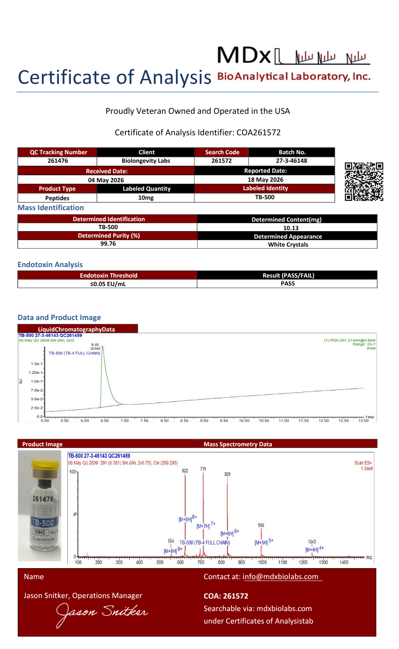
TB-500 Product Description
TB-500 is a synthetic version of Thymosin Beta-4, a naturally occurring 43 amino acid peptide found in virtually all mammalian cells. Thymosin Beta-4 acts as a multifunctional regulatory peptide comprising distinct bioactive regions within its structure: the N-terminal tetrapeptide (Ac-SDKP) mediates anti-inflammatory and antifibrotic activities, amino acids 1-15 inhibit apoptosis and promote cell survival, while the central actin-binding domain (amino acids 17-23, containing LKKTET) drives angiogenesis, cell migration, and wound healing.
Thymosin beta-4 is among the most abundant beta-thymosins found in mammalian tissue and is largely unstructured in aqueous solution. That conformational flexibility is central to how it is studied: an unfolded backbone can recognize multiple molecular targets, which is the working explanation for the wide range of binding partners reported for it.
Two sequence regions do most of the mechanistic work in the literature. The central actin-binding motif KLKKTET, spanning residues 17 through 23, mediates G-actin sequestration. The N-terminal tetrapeptide AcSDKP is a separately characterized fragment with its own experimental record.
Compound Information
| Property |
Value |
| Synonyms |
Thymosin Beta-4; Thymosin β4; Tβ4; TB4; Timbetasin (INN); TMSB4X gene product |
| CAS Number |
77591-33-4 |
| PubChem CID |
45382195 |
| Molecular Formula |
C₂₁₂H₃₅₀N₅₆O₇₈S |
| Molecular Weight |
4963.44 g/mol |
| Amino Acid Sequence |
Ac-SDKPDMAEIEKFDKSKLKKTETQEKNPLPSKETIEQEKQAGES |
| Sequence Length |
43 residues, N-terminally acetylated |
| Actin-Binding Motif |
KLKKTET (residues 17 to 23) |
| Source |
Synthetic |
| InChIKey |
UGPMCIBIHRSCBV-XNBOLLIBSA-N |
| Purity |
≥99% (HPLC) |
| Appearance |
Lyophilized white powder |
| Solubility |
Soluble in water |
| Storage |
-20°C, protect from light |
TB-500 (Thymosin Beta-4) Peptide Structure
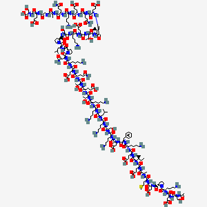
Source: PubChem
Storage and Handling
- Store the lyophilized compound at -20°C, sealed and protected from light.
- After reconstitution, store at 2°C to 8°C and use promptly.
- Allow the vial to reach room temperature before opening to limit moisture uptake.
- Brief sonication may be required for complete dissolution at higher working concentrations.
- Maintain aseptic handling to preserve compound integrity.
Lyophilized Format
This compound ships in lyophilized (freeze-dried) form. Freeze-drying supports long-term storage stability and preserves compound integrity. No fillers are added.
TB-500 Research
The defining molecular property of thymosin beta-4 is actin monomer sequestration. The peptide binds G-actin and inhibits its polymerization into F-actin, and structural analysis indicates the molecule is largely or completely unfolded in solution, a feature that has been proposed to explain the breadth of ligand recognition reported for it [1]. Work in cerebral endothelial cell culture has recorded a corresponding shift in the F-actin to G-actin ratio alongside changes in tight junction protein expression [2].
Endothelial migration has been characterized in detail. Using mutants deprived of the N-terminal AcSDKP tetrapeptide, of the KLKKTET actin-binding sequence, and carrying a Lys16Ala point mutation, one study showed that increased intracellular expression of the peptide was sufficient to induce PAI-1 gene expression in endothelial cells and to stimulate expression and release of MMP-1, MMP-2, and MMP-3 [3]. Notably, the transition of endothelial cells from a quiescent to a proangiogenic phenotype did not require the AcSDKP sequence and depended only partially on G-actin binding.
Angiogenic signaling has been mapped to specific pathways. In endothelial culture and a limb ischemia model, peptide overexpression was associated with changes in angiopoietin-2, Tie2, and VEGF-A expression, and pathway inhibitor work implicated Notch and NF-kappaB signaling in the response [4]. Cardiac research models have examined the peptide’s antifibrotic and proangiogenic activity, with the SRF-MRTF-G-actin transcriptional pathway identified as an indirect route by which it influences cellular motility [5].
Additional experimental literatures cover endothelial cell viability and senescence in induced pluripotent stem cell-derived cultures [6], hepatic stellate cell proliferation and migration in fibrosis models [7], and corneal epithelial models following alkali injury, where re-epithelialization rates and inflammatory cytokine transcript levels were measured [8]. Tumor cell migration studies have examined the peptide’s interaction with hypoxia-inducible factor-1 alpha [9].
Why Sequence Fragment Matters in Study Design
Because the literature contains data on the full 43-residue peptide, on the isolated KLKKTET motif, and on the AcSDKP tetrapeptide, results are not interchangeable across those forms. The mutant work described above showed directly that some observed responses persist when the actin-binding sequence is disabled and others do not [3]. Laboratories should confirm which construct a cited study used before designing a comparison. Quantification is a related concern, and published reviews have documented substantial variability in reported circulating concentrations traced to sample preparation and assay specificity [10].
| Research Area |
In Vitro Application |
| Cytoskeletal regulation |
G-actin sequestration and F-actin to G-actin ratio assays |
| Endothelial cell biology |
Migration, tube formation, and PAI-1 and MMP expression assays |
| Angiogenic signaling |
Notch, NF-kappaB, VEGF-A, and Tie2 pathway profiling |
| Barrier function |
Tight junction protein expression in endothelial monolayer models |
| Fibrosis research models |
Hepatic stellate cell proliferation and migration assays |
References
- Bubb MR. (2003). Thymosin beta 4 interactions. Vitamins and Hormones. https://doi.org/10.1016/S0083-6729(03)01008-2
- Song K, Han HJ, Kim S, Kwon J. (2019). Thymosin beta 4 attenuates PrP(106-126)-induced human brain endothelial cells dysfunction. European Journal of Pharmacology. https://doi.org/10.1016/j.ejphar.2019.172891
- Cierniewski CS, Malinowski M, Bednarek R, Cierniewska-Cieslak A. (2007). Adhesive and proteolytic phenotype of migrating endothelial cells induced by thymosin beta-4. Annals of the New York Academy of Sciences. https://doi.org/10.1196/annals.1415.019
- Lv S, Cai H, Xu Y, Dai J, Rong X, Zheng L. (2020). Thymosin-beta 4 induces angiogenesis in critical limb ischemia mice via regulating Notch/NF-kappaB pathway. International Journal of Molecular Medicine. https://pmc.ncbi.nlm.nih.gov/articles/PMC7447324/
- Pipes GT, Yang J. (2016). Cardioprotection by Thymosin Beta 4. Vitamins and Hormones. https://doi.org/10.1016/bs.vh.2016.04.004
- Su L, Kong X, Loo S, et al. (2022). Thymosin beta-4 improves endothelial function and reparative potency of diabetic endothelial cells differentiated from patient induced pluripotent stem cells. Stem Cell Research & Therapy. https://pmc.ncbi.nlm.nih.gov/articles/PMC8751378/
- Kim J, Jung Y. (2015). Potential role of thymosin Beta 4 in liver fibrosis. International Journal of Molecular Sciences. https://pmc.ncbi.nlm.nih.gov/articles/PMC4463665/
- Sosne G, Szliter EA, Barrett R, Kernacki KA, Kleinman H, Hazlett LD. (2002). Thymosin beta 4 promotes corneal wound healing and decreases inflammation in vivo following alkali injury. Experimental Eye Research. https://doi.org/10.1006/exer.2001.1125
- Ryu YK, Im YS, Moon EY. (2010). Cooperation of actin-sequestering protein, thymosin beta-4 and hypoxia inducible factor-1 alpha in tumor cell migration. Oncology Reports. https://doi.org/10.3892/or_00000997
- Tan WKY, Purnamawati K, Pakkiri LS, Tan SH, Yang X, Chan MY, Drum CL. (2018). Sources of variability in quantifying circulating thymosin beta-4: literature review and recommendations. Expert Opinion on Biological Therapy. https://doi.org/10.1080/14712598.2018.1448382