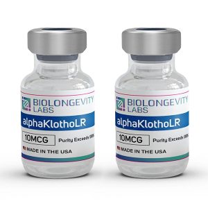
Product Description
alphaKlothoLR is a patented, long-release version of the recombinant Klotho protein, which contains specific amino acid sequences, linkers, and an albumin binding group that allows for a sustained delivery of Klotho with cellular activity up to 19 days.
This research-grade construct combines α-Klotho protein with a proprietary 29-amino acid albumin binding peptide (GGSGGSGGSGGRLIEDICLPRWGCLWEDD). The modification enables strong albumin binding affinity (<20 nM) while maintaining native FGF23 binding activity (15-30 nM).
Produced through recombinant expression with rigorous third-party testing. Each batch includes HPLC and LC-MS verification to confirm molecular identity and purity standards. Complete analytical documentation supports laboratory applications investigating protein-albumin interactions, FGF23 signaling pathways, and albumin binding mechanisms.
For in vitro research use only.
Klotho Protein Information
| Property |
Value |
| Peptide Sequence |
α-Klotho protein (1012 amino acids) + albumin binding construct: GGSGGSGGSGGRLIEDICLPRWGCLWEDD |
| Molecular Weight |
~133-135 kDa (full construct with albumin binder) |
| Albumin Binding Affinity (Kd) |
<20 nM |
| FGF23 Binding Affinity (Kd) |
15-30 nM (optimized: 16.2 nM) |
| Synonyms |
α-Klotho-albumin binding construct, Modified Klotho with glycine-serine linker |
Lyophilized Peptides:
These peptides are freeze-dried, a process that not only extends shelf life but also preserves the purity and integrity of the peptides during storage. We do not use any fillers in this process.
Product Usage:
This PRODUCT IS INTENDED AS A RESEARCH CHEMICAL ONLY. This designation allows the use of research chemicals strictly for in vitro testing and laboratory experimentation only. This product should only be handled by licensed, qualified professionals. This product is not a drug, food, or cosmetic and may not be misbranded, misused or mislabeled as a drug.
Klotho Research
Research on α-Klotho spans multiple systems, revealing its role as both a membrane-bound coreceptor and a circulating protein with diverse enzymatic activities.
Mineral Metabolism and Bone-Kidney Interactions
Klotho functions as an obligate coreceptor for FGF23, creating a complex that regulates phosphate excretion by the kidneys[1]. The protein also modulates sodium-phosphate cotransporters in kidney tubules through its enzymatic activity, affecting how cells reabsorb or excrete phosphate[2].
Beyond the FGF23 pathway, Klotho influences vitamin D metabolism and parathyroid hormone secretion[3]. These mechanisms connect mineral balance across bone and kidney tissue.
Oxidative Stress and Cellular Defense
Klotho activates Nrf2, a master regulator of antioxidant gene expression. This activation increases production of protective enzymes including heme oxygenase-1 and glutathione-related enzymes[4].
The protein also works through FoxO transcription factors to regulate manganese superoxide dismutase expression. This coordination creates cellular responses that reduce reactive oxygen species accumulation in cardiac, lung, vascular, and kidney tissue[5].
Cardiovascular and Vascular Function
In cardiac cells, Klotho modulates calcium channels and ionic currents through PI3K/Akt signaling pathways[6]. The protein affects vascular smooth muscle cells by inducing antioxidant defenses that protect against vascular aging processes[7].
Klotho also influences nitric oxide signaling in endothelial cells and demonstrates protective effects against vascular calcification and fibrosis[8].
Neurological Function and Synaptic Activity
Research shows Klotho affects synaptic transmission and plasticity in hippocampal circuits[9]. In astrocytes, the protein enhances glycolytic metabolism, potentially supporting neuronal energy supply.
Klotho provides protection against neurotoxic insults through oxidative stress reduction and PI3K/Akt pathway regulation. The protein also modulates autophagy and endoplasmic reticulum stress responses in neuronal tissues[10].
Inflammatory Response Modulation
Klotho inhibits NF-κB, a key regulator of inflammatory gene expression, reducing production of inflammatory cytokines and chemokines[4]. The protein also affects MAPK pathways that transmit inflammation and stress signals[11].
In various injury models, Klotho reduces inflammatory cell infiltration and decreases pro-inflammatory molecule expression.
Glucose Metabolism and Insulin Signaling
Klotho’s relationship with insulin signaling involves context-dependent mechanisms[12]. In pancreatic beta cells, the protein enhances glucose-stimulated insulin secretion through calcium channel regulation[13].
In liver tissue, soluble Klotho improves glucose and lipid metabolism by modulating the IGF1R/PI3K/Akt/mTOR pathway[14]. The protein influences cell surface expression of TRPV2 channels, which facilitate calcium entry.
Skeletal Muscle and Regeneration
Klotho participates in muscle progenitor cell function and their progression through regenerative stages[15]. When Klotho levels decline, muscle progenitor cells exhibit mitochondrial DNA damage and reduced cellular energy production[16].
Research documents correlations between circulating Klotho levels and grip strength, suggesting connections between the protein and functional muscle capacity[17].
Wnt and TGF-β Signaling Pathways
Klotho functions as an antagonist of Wnt signaling by binding to Wnt ligands and blocking their interaction with cell surface receptors[18]. This inhibition affects cell proliferation, differentiation, and tissue development programs.
The protein also interacts with TGF-β signaling pathways, which regulate fibrosis, inflammation, and cellular senescence in various tissues[19].
FoxO Activation and Stress Resistance
Klotho activates FoxO transcription factors by inhibiting the PI3K/Akt pathway[20]. This allows FoxO proteins to translocate to the nucleus and induce expression of protective genes.
Target genes include those encoding antioxidant enzymes, DNA repair proteins, and autophagy regulators[21]. This signaling axis links Klotho to cellular stress responses and functional maintenance.
Mitochondrial Function and Energetics
Klotho influences mitochondrial biogenesis and the generation of new mitochondria[20]. In muscle progenitor cells, Klotho deficiency leads to mitochondrial DNA damage and impaired respiratory function[16].
The protein modulates expression of mitochondrial uncoupling proteins and regulators of mitochondrial dynamics, affecting cellular metabolic programs[22].
DNA Integrity and Genomic Protection
Research indicates Klotho preserves mitochondrial DNA integrity when cells face oxidative stress[10]. The protein reduces DNA double-strand breaks caused by various insults[23].
These protective effects involve Akt signaling and possible DNA repair pathway activation, connecting to broader roles in cellular senescence processes.
Ion Channel and Membrane Transport Regulation
Klotho functions as a glycosidase that removes sugar molecules from membrane proteins[24]. For the TRPV5 calcium channel, Klotho removes sialic acid residues from N-glycans, affecting channel trafficking and stability[25].
This modification prevents channel endocytosis, increasing surface expression and enhancing calcium reabsorption in kidney tubules[26].
Autophagy and Protein Quality Control
Klotho influences autophagic flux and interacts with endoplasmic reticulum stress pathways[9]. In neurons, the protein increases proteasome activity, clearing damaged proteins[10].
This coordination between Klotho and cellular clearance mechanisms may be particularly relevant in tissues where protein aggregation occurs.
Cell Survival and Apoptosis Regulation
Klotho modulates programmed cell death through effects on the PI3K/Akt pathway and downstream caspase enzymes[27]. The protein affects expression of pro-apoptotic and anti-apoptotic proteins[23].
In various cell types, Klotho protects against cytotoxic insults and reduces apoptosis induced by oxidative stress or inflammatory signals[11].
Anti-Fibrotic Properties
Klotho demonstrates anti-fibrotic properties by modulating TGF-β signaling, a major driver of fibrosis in kidney, liver, and lung tissue[28]. Through Wnt signaling effects, Klotho influences epithelial-to-mesenchymal transition[18].
The protein reduces expression of profibrotic genes and extracellular matrix proteins in various organ injury models[19].
Enzymatic Activities
Klotho possesses β-glucuronidase activity, capable of cleaving glucuronic acid residues from substrates[29]. The protein also functions as a sialidase, removing sialic acid from glycoproteins[30].
These enzymatic activities occur through specific catalytic sites in Klotho’s extracellular domain, allowing direct modification of membrane protein function[31].
References
- M. S. Razzaque, “The FGF23–Klotho axis: endocrine regulation of phosphate homeostasis,” Springer Science and Business Media LLC, Nov. 2009. doi: 10.1038/nrendo.2009.196. https://doi.org/10.1038/nrendo.2009.196
- M. C. Hu et al., “Klotho: a novel phosphaturic substance acting as an autocrine enzyme in the renal proximal tubule,” Wiley, May 2010. doi: 10.1096/fj.10-154765. https://doi.org/10.1096/fj.10-154765
- M. C. Hu, K. Shiizaki, M. Kuro-o, and O. W. Moe, “Fibroblast Growth Factor 23 and Klotho: Physiology and Pathophysiology of an Endocrine Network of Mineral Metabolism,” Annual Reviews, Feb. 2013. doi: 10.1146/annurev-physiol-030212-183727. https://doi.org/10.1146/annurev-physiol-030212-183727
- X. Zhao et al., “New insights into the role of Klotho in inflammation and fibrosis: molecular and cellular mechanisms,” Frontiers Media SA, Sep. 2024. doi: 10.3389/fimmu.2024.1454142. https://doi.org/10.3389/fimmu.2024.1454142
- M. Yamamoto et al., “Regulation of Oxidative Stress by the Anti-aging Hormone Klotho*♦,” Elsevier BV, Nov. 2005. doi: 10.1074/jbc.m509039200. https://doi.org/10.1074/jbc.m509039200
- Y. Hung et al., “Klotho modulates electrical activity and calcium homeostasis in pulmonary vein cardiomyocytes via PI3K/Akt signalling,” Oxford University Press (OUP), Jun. 2020. doi: 10.1093/europace/euaa100. https://doi.org/10.1093/europace/euaa100
- G. Maltese et al., “The anti‐ageing hormone klotho induces Nrf2‐mediated antioxidant defences in human aortic smooth muscle cells,” Wiley, Oct. 2016. doi: 10.1111/jcmm.12996. https://doi.org/10.1111/jcmm.12996
- C. B. Leibrock et al., “NH4Cl Treatment Prevents Tissue Calcification in Klotho Deficiency,” Ovid Technologies (Wolters Kluwer Health), Oct. 2015. doi: 10.1681/asn.2014030230. https://doi.org/10.1681/asn.2014030230
- A. M. Orellana, C. H. Mazucanti, L. P. dos Anjos, L. de Sá Lima, E. M. Kawamoto, and C. Scavone, “Klotho increases antioxidant defenses in astrocytes and ubiquitin–proteasome activity in neurons,” Springer Science and Business Media LLC, Sep. 2023. doi: 10.1038/s41598-023-41166-6. https://doi.org/10.1038/s41598-023-41166-6
- J. Mytych, “Klotho and neurons: mutual crosstalk between autophagy, endoplasmic reticulum, and inflammatory response,” Medknow, 2021. doi: 10.4103/1673-5374.303014. https://doi.org/10.4103/1673-5374.303014
- A. Al-Kadi, A. Anter, R. R. Rofaeil, M. M. Sayed-Ahmed, and A.-S. F. Ahmed, “Klotho: A multifaceted protector in sepsis-induced organ damage and a potential therapeutic target,” Baishideng Publishing Group Inc., Sep. 2025. doi: 10.5492/wjccm.v14.i3.103458. https://doi.org/10.5492/wjccm.v14.i3.103458
- T. Landry, D. Shookster, and H. Huang, “Circulating α-klotho regulates metabolism via distinct central and peripheral mechanisms,” Elsevier BV, Aug. 2021. doi: 10.1016/j.metabol.2021.154819. https://doi.org/10.1016/j.metabol.2021.154819
- R. Anour, O. Andrukhova, E. Ritter, U. Zeitz, and R. G. Erben, “Klotho Lacks a Vitamin D Independent Physiological Role in Glucose Homeostasis, Bone Turnover, and Steady-State PTH Secretion In Vivo,” Public Library of Science (PLoS), Feb. 2012. doi: 10.1371/journal.pone.0031376. https://doi.org/10.1371/journal.pone.0031376
- Y. Lin and Z. Sun, “Antiaging Gene Klotho Enhances Glucose-Induced Insulin Secretion by Up-Regulating Plasma Membrane Levels of TRPV2 in MIN6 β-Cells,” The Endocrine Society, May 2012. doi: 10.1210/en.2012-1091. https://doi.org/10.1210/en.2012-1091
- Z. Clemens et al., “The biphasic and age-dependent impact of klotho on hallmarks of aging and skeletal muscle function,” eLife Sciences Publications, Ltd, Apr. 2021. doi: 10.7554/elife.61138. https://doi.org/10.7554/elife.61138
- E. Arroyo, A. D. Troutman, R. N. Moorthi, K. G. Avin, A. R. Coggan, and K. Lim, “Klotho: An Emerging Factor With Ergogenic Potential,” Frontiers Media SA, Jan. 2022. doi: 10.3389/fresc.2021.807123. https://doi.org/10.3389/fresc.2021.807123
- R. D. Semba et al., “Relationship of low plasma klotho with poor grip strength in older community-dwelling adults: the InCHIANTI study,” Springer Science and Business Media LLC, Jul. 2011. doi: 10.1007/s00421-011-2072-3. https://doi.org/10.1007/s00421-011-2072-3
- A. Kale, H. Sankrityayan, H.-J. Anders, and A. B. Gaikwad, “Klotho in kidney diseases: a crosstalk between the renin–angiotensin system and endoplasmic reticulum stress,” Oxford University Press (OUP), Nov. 2021. doi: 10.1093/ndt/gfab340. https://doi.org/10.1093/ndt/gfab340
- M. C. Hu, A. Bian, J. Neyra, and M. Zhan, “Klotho, stem cells, and aging,” Informa UK Limited, Aug. 2015. doi: 10.2147/cia.s84978. https://doi.org/10.2147/cia.s84978
- J. Donate-Correa, B. Martín-Carro, J. B. Cannata-Andía, C. Mora-Fernández, and J. F. Navarro-González, “Klotho, Oxidative Stress, and Mitochondrial Damage in Kidney Disease,” MDPI AG, Jan. 2023. doi: 10.3390/antiox12020239. https://doi.org/10.3390/antiox12020239
- X. Sun, W.-D. Chen, and Y.-D. Wang, “DAF-16/FOXO Transcription Factor in Aging and Longevity,” Frontiers Media SA, Aug. 2017. doi: 10.3389/fphar.2017.00548. https://doi.org/10.3389/fphar.2017.00548
- A. Sahu et al., “Age-related declines in α-Klotho drive progenitor cell mitochondrial dysfunction and impaired muscle regeneration,” Springer Science and Business Media LLC, Nov. 2018. doi: 10.1038/s41467-018-07253-3. https://doi.org/10.1038/s41467-018-07253-3
- S.-J. Kim et al., “Klotho, an antiaging molecule, attenuates oxidant-induced alveolar epithelial cell mtDNA damage and apoptosis,” American Physiological Society, Jul. 2017. doi: 10.1152/ajplung.00063.2017. https://doi.org/10.1152/ajplung.00063.2017
- S.-K. Cha, B. Ortega, H. Kurosu, K. P. Rosenblatt, M. Kuro-o, and C.-L. Huang, “Removal of sialic acid involving Klotho causes cell-surface retention of TRPV5 channel via binding to galectin-1,” Proceedings of the National Academy of Sciences, Jul. 2008. doi: 10.1073/pnas.0803223105. https://doi.org/10.1073/pnas.0803223105
- M. T. F. Wolf, S.-W. An, M. Nie, M. S. Bal, and C.-L. Huang, “Klotho Up-regulates Renal Calcium Channel Transient Receptor Potential Vanilloid 5 (TRPV5) by Intra- and Extracellular N-glycosylation-dependent Mechanisms,” Elsevier BV, Dec. 2014. doi: 10.1074/jbc.m114.616649. https://doi.org/10.1074/jbc.m114.616649
- G. D. Dalton, J. Xie, S.-W. An, and C.-L. Huang, “New Insights into the Mechanism of Action of Soluble Klotho,” Frontiers Media SA, Nov. 2017. doi: 10.3389/fendo.2017.00323. https://doi.org/10.3389/fendo.2017.00323
- P. Zhou, C. Zhao, Y. Chen, X. Liu, C. Wu, and Z. Hu, “Klotho activation of Nrf2 inhibits the ferroptosis signaling pathway to ameliorate sepsis-associated acute kidney injury,” AME Publishing Company, Dec. 2023. doi: 10.21037/tau-23-573. https://doi.org/10.21037/tau-23-573
- A. D. Hajare, N. Dagar, and A. B. Gaikwad, “Klotho antiaging protein: molecular mechanisms and therapeutic potential in diseases,” Springer Science and Business Media LLC, Mar. 2025. doi: 10.1186/s43556-025-00253-y. https://doi.org/10.1186/s43556-025-00253-y
- Y. Xu and Z. Sun, “Molecular Basis of Klotho: From Gene to Function in Aging,” The Endocrine Society, Feb. 2015. doi: 10.1210/er.2013-1079. https://doi.org/10.1210/er.2013-1079
- O. Tohyama et al., “Klotho is a novel beta-glucuronidase capable of hydrolyzing steroid beta-glucuronides.,” Journal of Biological Chemistry, vol. 279 11, pp. 9777–84, 2004.
- E. S. Kuzina et al., “Structures of ligand-occupied β-Klotho complexes reveal a molecular mechanism underlying endocrine FGF specificity and activity,” Proceedings of the National Academy of Sciences, Apr. 2019. doi: 10.1073/pnas.1822055116. https://doi.org/10.1073/pnas.1822055116
Certificate of Analysis (COA) for Every Batch
A Certificate of Analysis (COA) is a document that verifies a compound’s identity, purity, and batch quality through independent laboratory testing. Every compound from BioLongevity Labs ships with a COA tied to its specific batch, so researchers can confirm exactly what they received before it enters a protocol.
Each COA reports results from third-party laboratory analysis, including:
- High-performance liquid chromatography (HPLC) for purity, typically confirmed at 99% or higher
- Liquid chromatography mass spectrometry (LC-MS) for molecular identity and mass confirmation
- Sterility and endotoxin screening where applicable
- Chemical contaminant and residual solvent checks
COAs are sourced from independent certified labs rather than in-house testing alone, giving researchers a verifiable record of molecular integrity for each batch. All compounds are supplied for research use only.
Review the COAs for this batch below, or browse the full COA library.
Klotho (260086)
 (20mcg) 2")
Klotho (260797)
 (20mcg) 3")
×
![]()

 (20mcg) 1")