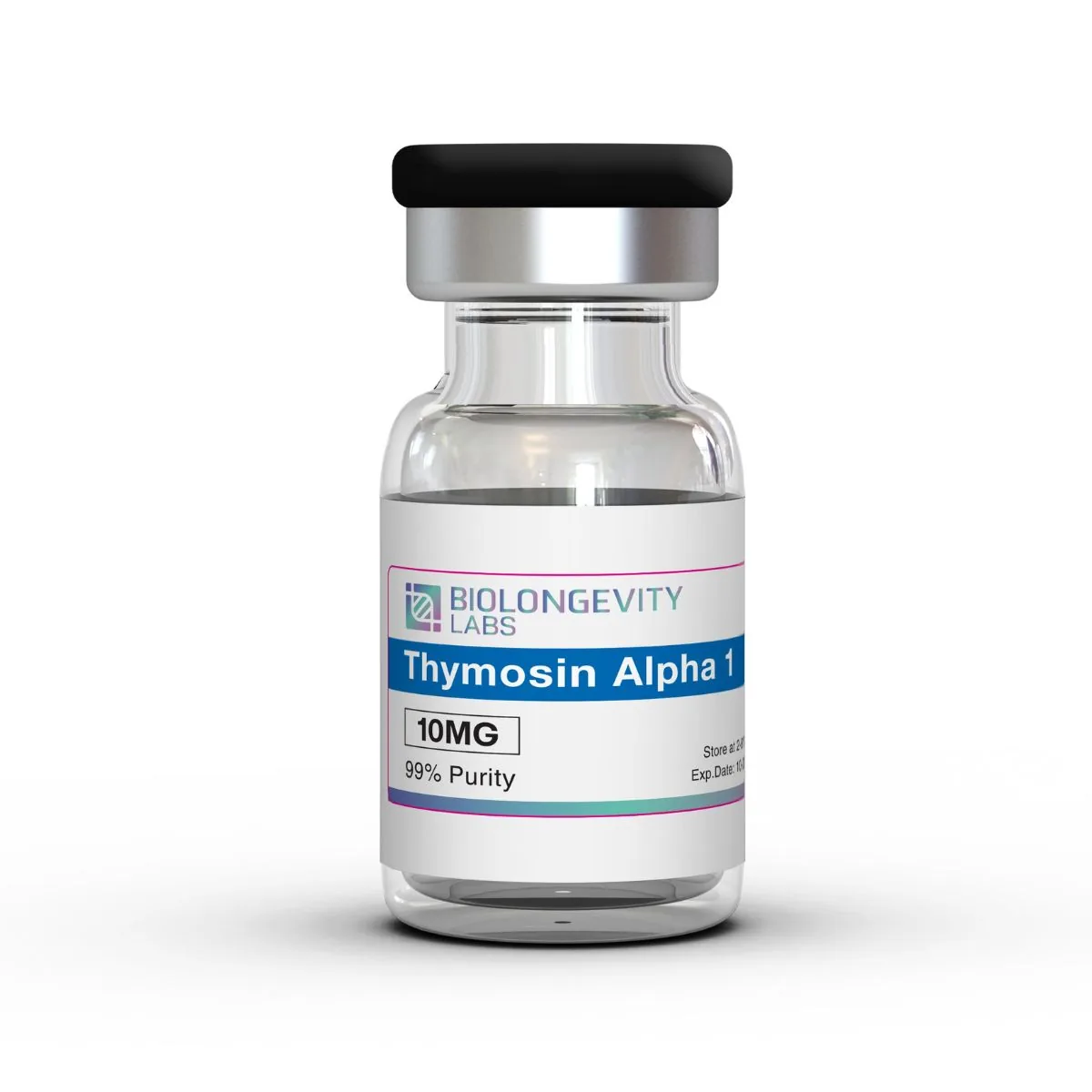
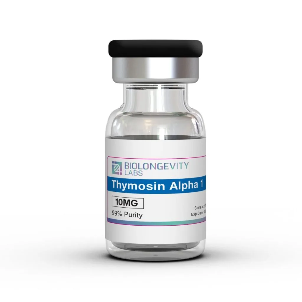
Thymosin alpha 1 (Tα1, thymalfasin) is a synthetic, N-terminally acetylated 28-residue peptide that matches the sequence first isolated from the thymosin fraction 5 extract of thymus tissue. BioLongevity Labs supplies it as 10mg of lyophilized white powder in a sealed vial, with ≥99% purity confirmed by HPLC. The peptide is characterized in laboratory research on Toll-like receptor (TLR2 and TLR9) signalling in dendritic cell and macrophage models. It is also studied for T-cell maturation markers and cytokine expression in cell-culture systems.
≥99% Purity
Third-Party Tested
COA Available
U.S. Manufactured
Quantity
Out of stock
Notify me when in stock
Third-Party Verified Quality
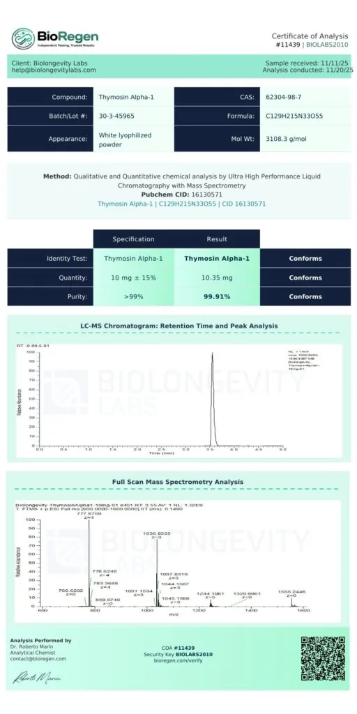
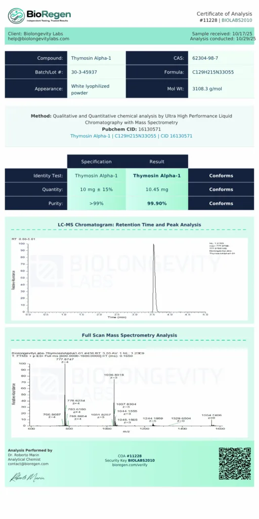
≥99% purity verified through independent UHPLC-MS testing. Certificates of Analysis are available before purchase.
COAs available before purchase
U.S.-manufactured research-grade
Same-business-day U.S. dispatch for in-stock orders placed before 12 PM PT
Secure Checkout · Free shipping on orders over $400
Thymosin alpha 1 is the N-terminal fragment of prothymosin alpha, a larger nuclear protein. In tissue, a lysosomal asparaginyl endopeptidase (legumain) cleaves prothymosin alpha between Asn28 and Gly29 to release the 28-residue peptide.
The sequence is highly acidic, with a run of aspartate and glutamate residues and an acetylated serine at the N-terminus. This research peptide is produced by solid-phase peptide synthesis, which gives a defined sequence with no tissue-derived material.
In the laboratory, the TA1 peptide is used as a reference ligand for innate signalling studies. Typical work covers dendritic cell maturation, macrophage MAPK activation, thymocyte surface binding and cytokine readouts in primary cell cultures.
Store the lyophilized compound at -20°C, protected from light and moisture.
After reconstitution, store at 2 to 8°C and use promptly.
Maintain aseptic handling to preserve compound integrity.
Lyophilized Format
This compound ships in lyophilized (freeze-dried) form. Freeze-drying supports long-term storage stability and preserves compound integrity. No fillers are added.
Research Use Disclaimer
Thymosin Alpha 1 is supplied for research use only. It is not a drug, food, cosmetic, or dietary supplement and has not been evaluated by the FDA. By purchasing, the buyer confirms the compound will be used solely for in vitro research.
Frequently Asked Questions
What Is Thymosin Alpha 1?
Thymosin alpha 1 is an N-terminally acetylated 28-residue peptide derived from prothymosin alpha and first isolated from thymus tissue. It is studied in laboratory models of Toll-like receptor signalling, dendritic cell maturation and cytokine expression.
What Is the CAS Number of Thymosin Alpha 1?
The CAS number of thymosin alpha 1 is 62304-98-7, and its PubChem CID is 16130571. The peptide has the molecular formula C₁₂₉H₂₁₅N₃₃O₅₅ and a molecular weight of 3108.3 g/mol.
Is Thymosin Alpha 1 the Same as TB-500?
No. Thymosin alpha 1 is a 28-residue peptide cut from prothymosin alpha, while TB-500 is a synthetic peptide based on thymosin beta-4, a separate protein with an unrelated sequence.
How Should Thymosin Alpha 1 Be Stored?
Lyophilized thymosin alpha 1 should be stored at -20°C, protected from light and moisture. After reconstitution, keep the solution at 2 to 8°C and use it promptly.
How Is the Purity of Thymosin Alpha 1 Verified?
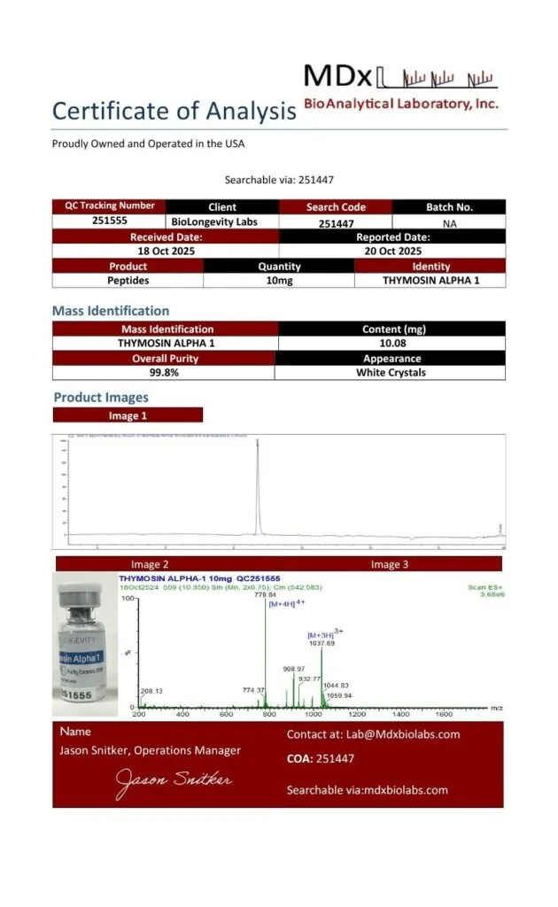
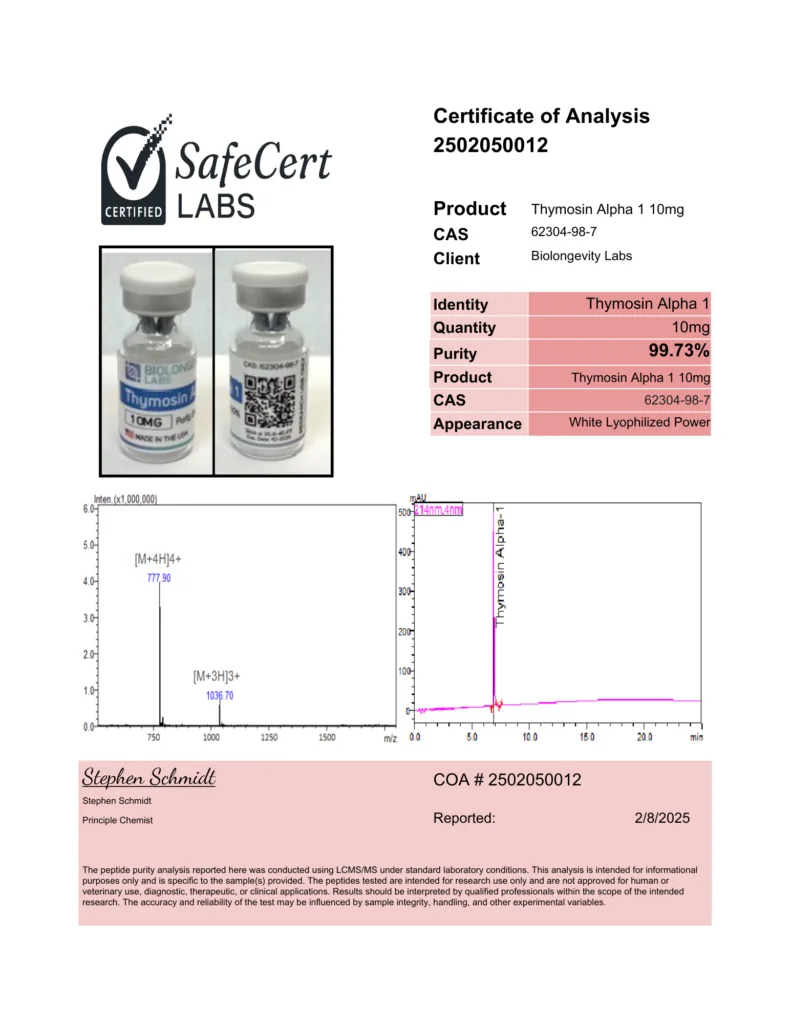
Thymosin alpha 1 purity of ≥99% is confirmed by HPLC at an independent third-party lab. The Certificate of Analysis is published on the product page and can be read before ordering.
Research Overview
Thymosin alpha 1 was first sequenced from calf thymus in 1977 as a heat-stable, highly acidic 28-residue component of thymosin fraction 5 [1]. Later work showed that the peptide is a natural cleavage product of prothymosin alpha, generated by the lysosomal enzyme legumain and present free in the cytosol of diverse mammalian tissues [2].
Today the peptide is made by solid-phase synthesis, and recombinant expression systems have also been described. In vitro assays with these preparations measure cytokine secretion and lymphocyte proliferation, and the peptide blocks steroid-induced apoptosis of thymocytes in culture [3].
The research threads below cover Toll-like receptor signalling in dendritic cells and macrophages, then T-cell maturation markers.
Toll-Like Receptor Signalling in Dendritic Cells
In dendritic cell models, Tα1 induced functional maturation and interleukin-12 production through the p38 MAPK and NF-κB pathway, routed through MyD88-dependent Toll-like receptor signalling [4]. Further work linked this activity to TLR9: Tα1 induced indoleamine 2,3-dioxygenase in dendritic cells, which required TLR9 and type I interferon receptor signalling and led to IL-10 production and regulatory T-cell generation [5].
The response depends on the co-stimulus. In monocyte-derived dendritic cells exposed to TLR3 and TLR7/8 agonists, Tα1 raised MHC class I and II expression and IL-6, TNF-α and IL-8 secretion. With TLR2 and TLR4 agonists, it lowered the same readouts [6].
Macrophage Signalling and Cytokine Output
In murine bone marrow-derived macrophages, Tα1 at 100 ng/mL activated p42/44 MAPK within 5 to 15 minutes. The MAPK inhibitor PD98059 blocked the resulting nitric oxide and TNF-α production in a concentration-dependent manner [7].
T-Cell Maturation Markers
Thymosin alpha 1 acts in vitro on T-cell precursors and induces surface markers. Immunofluorescence work found specific binding sites on immature, peanut agglutinin-positive (PNA+) thymocytes [8].
Research Area
In Vitro Application
TLR signalling
MyD88, p38 MAPK and NF-κB pathway activation in dendritic cell cultures
TLR9 and tolerance pathways
Indoleamine 2,3-dioxygenase induction and IL-10 readouts in dendritic cells
Co-stimulus studies
Comparing TLR2/4 and TLR3/7/8 agonist responses in monocyte-derived dendritic cells
Macrophage signalling
p42/44 MAPK phosphorylation, nitric oxide and TNF-α output in bone marrow-derived macrophages
T-cell maturation
Surface marker induction and binding studies in immature thymocyte fractions
Cytokine expression
IL-6, IL-8, IL-12 and TNF-α measurement in primary cell cultures
References
Goldstein AL, Low TL, McAdoo M, et al. (1977). Thymosin alpha1: isolation and sequence analysis of an immunologically active thymic polypeptide. Proceedings of the National Academy of Sciences of the United States of America. https://doi.org/10.1073/pnas.74.2.725
Sarandeses CS, Covelo G, Díaz-Jullien C, Freire M. (2003). Prothymosin alpha is processed to thymosin alpha 1 and thymosin alpha 11 by a lysosomal asparaginyl endopeptidase. The Journal of Biological Chemistry. https://doi.org/10.1074/jbc.M213005200
Romani L, Bistoni F, Gaziano R, et al. (2004). Thymosin alpha 1 activates dendritic cells for antifungal Th1 resistance through toll-like receptor signaling. Blood. https://doi.org/10.1182/blood-2003-11-4036
Romani L, Bistoni F, Perruccio K, et al. (2006). Thymosin alpha1 activates dendritic cell tryptophan catabolism and establishes a regulatory environment for balance of inflammation and tolerance. Blood. https://doi.org/10.1182/blood-2006-02-004762
Giacomini E, Severa M, Cruciani M, et al. (2015). Dual effect of Thymosin α 1 on human monocyte-derived dendritic cell in vitro stimulated with viral and bacterial toll-like receptor agonists. Expert Opinion on Biological Therapy. https://doi.org/10.1517/14712598.2015.1019460
Sodhi A, Paul S. (2002). Involvement of mitogen-activated protein kinases in the signal transduction pathway of bone marrow-derived macrophage activation in response to in vitro treatment with thymosin alpha 1. International Immunopharmacology. https://doi.org/10.1016/s1567-5769(01)00139-4
A Certificate of Analysis (COA) is a document that verifies a compound’s identity, purity and quality through independent laboratory testing. Every compound from BioLongevity Labs is third-party tested, and its COA is published here, so researchers can review the testing record before a compound enters a protocol.
Each COA reports results from third-party laboratory analysis, including:
Ultra-high-performance liquid chromatography with mass spectrometry (UHPLC-MS) for purity, measured against a 98% or 99% specification depending on the compound
Mass identification for molecular confirmation and content quantitation
Endotoxin testing by Limulus amebocyte lysate (LAL) or recombinant Factor C (rFC) assay, depending on the laboratory
Visual and physical characterization of the finished material
How to Verify a COA Independently
You can check every certificate against the issuing laboratory’s own records, not just the copy hosted here. Verification does not depend on BioLongevity Labs.
AV Analytics certificates carry a search code. Scan the QR code on the certificate to open the laboratory’s verification page, or enter the search code at avanalytical.com/coa-search to pull the official record.
BioRegen reports of analysis carry a Report ID and a Validation Key. Scan the QR code on the certificate to open the official record, or quote both identifiers when you contact the laboratory at the address printed on the report.
SafeCert Labs issued a number of earlier certificates. Each one carries a COA number and the signature of the reporting chemist. Quote that number when you ask the laboratory to confirm it.
Older certificates from other laboratories list their own verification details on the document.
When a batch is sent to more than one laboratory, each certificate resolves at its own issuing laboratory. A researcher can then check the results against two unrelated sources.
These COAs come from independent certified laboratories, not in-house testing. That gives researchers a verifiable record of molecular integrity for each compound. All compounds are supplied for research use only.
Scientifically reviewed by Dr. Ky H. Le, MD. Dr. Le is a board-certified family medicine physician with over 20 years of clinical experience. Dr. Le validates the scientific accuracy of all technical content and research citations.
Disclaimer: For Research Purposes Only
This content is provided strictly for research purposes and does not constitute an endorsement or recommendation for the non-laboratory application or improper handling of peptides designed for research. The information, including discussions about specific peptides and their researched benefits, is presented for informational purposes only and must not be construed as health, clinical, or legal guidance, nor an encouragement for non-research use. Peptides described here are solely for use in structured scientific study by authorized individuals. We advise consulting with research experts, medical practitioners, or legal counsel prior to any decisions about obtaining or utilizing these peptides. The expectation of responsible, ethical utilization of this information for legitimate investigative and scholarly objectives is paramount. This notice is dynamic and governs all provided content on research peptides.