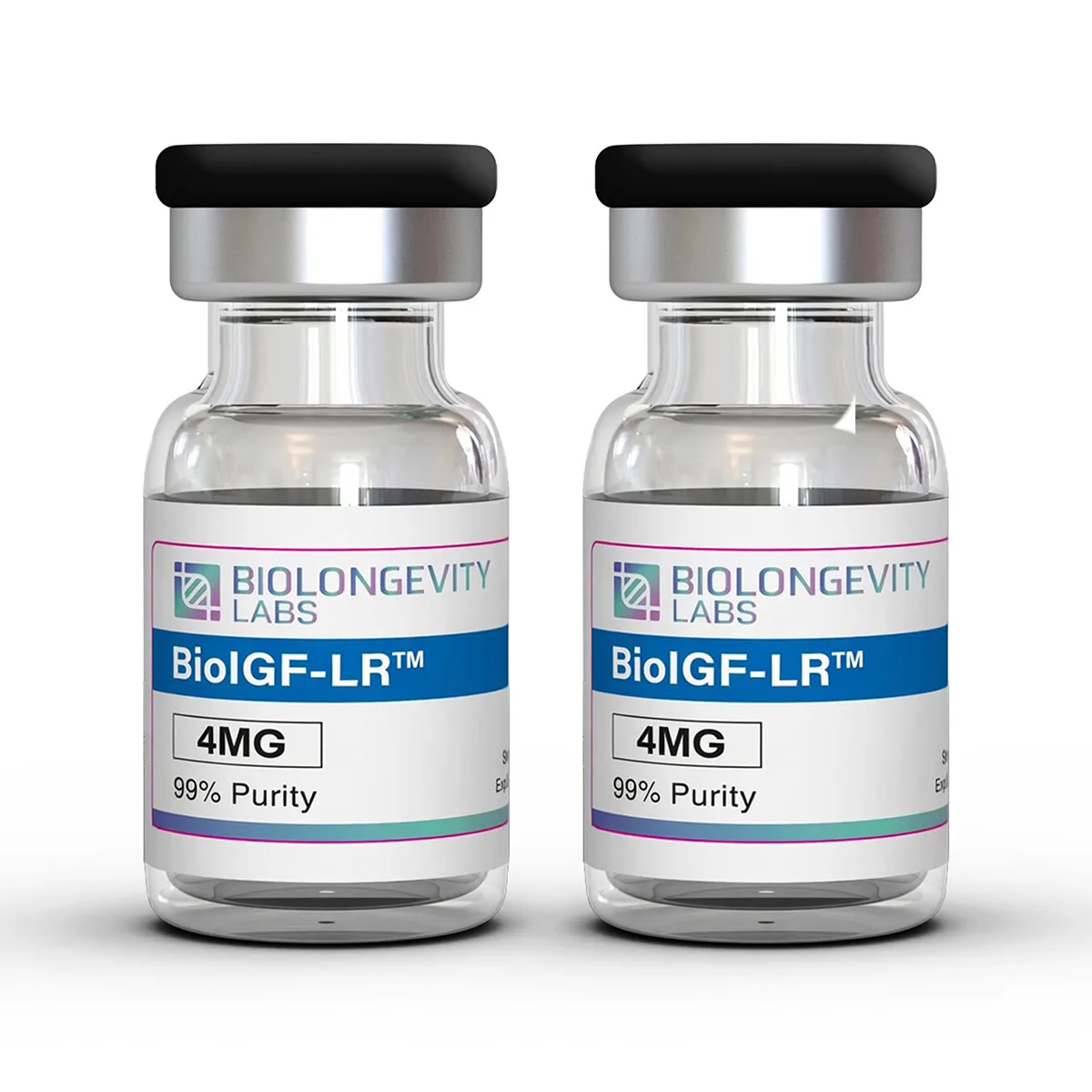
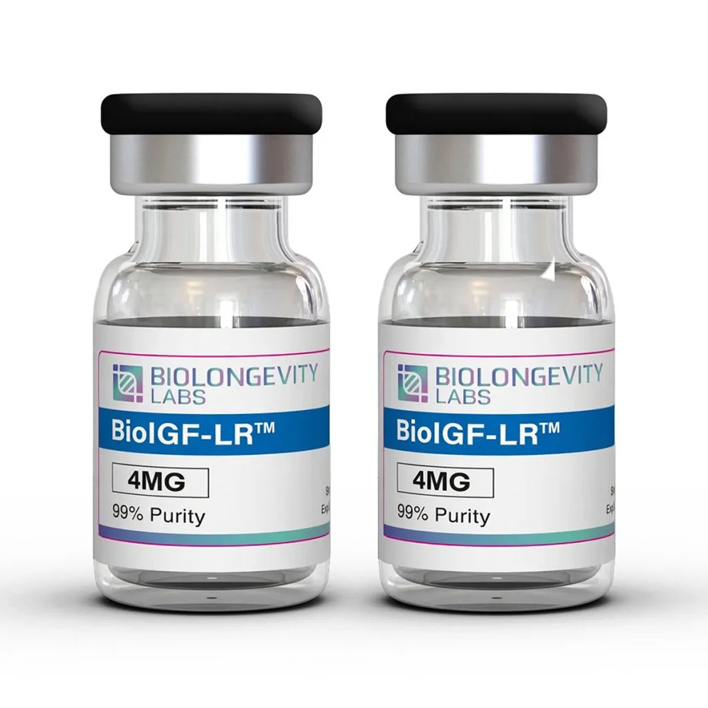
BioIGF-LR Product Description
IGF-1 is a signaling polypeptide studied across muscle, skeletal, metabolic, and neural research models. Its receptor, IGF-1R, activates the PI3K/Akt and MAPK cascades that laboratories use as readouts for cell proliferation, protein synthesis, and metabolic activity.
Native IGF-1 carries a short serum stability period, which limits the length of exposure achievable in longer experimental designs. BioIGF-LR is a modified-analog approach to that limitation. Structural changes of this kind are a standard strategy in peptide modification work, where sequence or terminal changes alter binding-protein affinity and molecular persistence.
Circulating IGF-1 concentrations decline across adult study populations with advancing age, which is one reason IGF-1 signaling remains an active area of laboratory investigation.
Compound Information
| Property |
Value |
| Compound Class |
Modified insulin-like growth factor 1 analog |
| Reported Serum Stability Period |
Approximately 17 days |
| Purity |
≥99% (HPLC) |
| Appearance |
Lyophilized white powder |
| Storage |
-20°C, protect from light |
Storage and Handling
- Store the lyophilized compound at 2°C to 8°C, protected from light.
- After reconstitution, store at 2°C to 8°C and use promptly.
- Avoid repeated freeze-thaw cycles, which degrade protein-class compounds.
- Maintain aseptic handling to preserve compound integrity.
Lyophilized Format
This compound ships in lyophilized (freeze-dried) form. Freeze-drying supports long-term storage stability and preserves compound integrity. No fillers are added.
Research Use Disclaimer
The compound is supplied for research use only. It is not a drug, food, cosmetic, or dietary supplement, has not been evaluated by the FDA, and is for research use only. By purchasing, the buyer confirms the compound will be used solely for in vitro research.
IGF-1 Research
IGF-1 signaling is one of the more closely mapped pathways in skeletal muscle research. Reduced IGF-1 signaling has been associated with myofibre atrophy in aging models, alongside changes in activity, nutrition, and motor neuron loss [1]. Reviews of the IGF-1/Akt/mTOR cascade describe it as the primary link between mechanical loading and muscle protein synthesis, and identify age-related dysregulation of that cascade as a research target [2].
Fat metabolism and skeletal muscle anabolic pathways run through overlapping signaling routes. Both involve PI3K/Akt and MAPK/ERK activation downstream of receptor tyrosine kinase activity, which is why IGF-1R occupancy is a shared readout across adipose and myogenic models [3].
At the cellular level, IGF-1 is associated with muscle satellite cell proliferation and myogenic differentiation, with IGF-1R activation of PI3K/Akt reported as the operative step in myoblast proliferation models [3]. IGF-1 signaling has also been mapped onto bone research, where it interacts with parathyroid hormone signaling and integrin mechanotransduction pathways in osteogenesis models [4].
Metabolic Research Findings
Reviews of IGF-1 deficiency describe associations with altered lipid profiles, insulin resistance, and glucose handling across animal and observational work [5]. Separate review work has characterized the short-term metabolic action of IGF-1 as insulin-like and the long-term action as growth-factor-like, with outcomes that vary by tissue and by concentration [6].
The concentration dependence runs in both directions. IGF1 expression was increased in adipose tissue in obesity models, and genetic down-regulation of IGF1 normalized lipogenic gene expression and energy metabolism in a diet-induced rodent model [7].
Cognitive and Neuronal Research Findings
IGF-1 receptor activity has been documented on both neurons and neuroglia, with reported involvement in myelination, synaptogenesis, and astrocytic homeostatic function [8]. Reviews of IGF signaling in neurodegeneration models describe disrupted PI3K/Akt and MAPK signaling downstream of IGF-1R as a recurring feature, alongside altered brain energy metabolism [9].
Akt phosphorylation inactivates Glycogen Synthase Kinase-3 Beta (GSK-3β). In rodent models, IGF-1 application restored activity in the PI3K/Akt/GSK-3β axis and reversed changes in apoptotic and inflammatory markers [11].
Concentration matters here as well. An eight-year follow-up study in adult study populations reported that the highest IGF-1 quintile was associated with lower processing capacity and global cognition scores at follow-up, with the authors proposing that intermediate concentrations track with better cognitive scores rather than high ones [10].
Sleep and Circadian Signaling Research
Circulating IGF-1 follows a circadian pattern, and its entry into the brain is activity-dependent. Work in mice with disrupted IGF-1 receptor activity in orexin neurons reported lower hypothalamic orexin levels, altered electrocorticographic patterns with predominant slow wave activity, and reduced sleep onset latency [13].
Follow-on work from the same group reported that IGF-1 modulates orexin levels and hepatic expression of circadian entrainment genes in an orexin-dependent manner, positioning IGF-1R signaling at the junction between metabolic state and circadian activity [14].
Observational work has reported lower serum IGF-1 concentrations in adult study populations with chronic insomnia disorder, with concentrations correlating with polysomnography-measured slow wave sleep time [12]. Rodent sleep deprivation models reported down-regulated IGF-1 alongside reduced PI3K/Akt/GSK-3β activation in brain tissue [11].
Taken together, these findings point toward IGF-1 signaling being concentration-dependent and tissue-dependent, which is a reason laboratories value compounds with a defined and extended serum stability period.
| Research Area |
In Vitro Application |
| Myogenesis |
Myoblast proliferation and differentiation assays in C2C12 and L6 cell lines |
| Receptor signaling |
IGF-1R binding affinity and PI3K/Akt/mTOR phosphorylation readouts |
| MAPK pathway work |
ERK1/2 phosphorylation time-course assays across adipocyte and myotube models |
| Adipocyte metabolism |
Lipogenic gene expression profiling in cultured adipocytes |
| Skeletal models |
Osteoblast differentiation and mineralization assays |
| Neuronal models |
Neurite outgrowth and neurotrophic signaling assays |
| GSK-3β signaling |
Akt-dependent GSK-3β phosphorylation readouts in cultured neurons |
| Hypothalamic models |
IGF-1R expression profiling in orexin-expressing neuronal preparations |
| Analytical method development |
HPLC and LC-MS reference standard for analog stability work |
References
- Grounds MD (2002). Reasons for the degeneration of ageing skeletal muscle: a central role for IGF-1 signalling. Biogerontology 3(1-2):19-24. https://doi.org/10.1023/a:1015234709314
- Barclay RD, Burd NA, Tyler C, Tillin NA, Mackenzie RW (2019). The Role of the IGF-1 Signaling Cascade in Muscle Protein Synthesis and Anabolic Resistance in Aging Skeletal Muscle. Frontiers in Nutrition 6:146. https://doi.org/10.3389/fnut.2019.00146
- Ahmad SS, Ahmad K, Lee EJ, Lee Y-H, Choi I (2020). Implications of Insulin-Like Growth Factor-1 in Skeletal Muscle and Various Diseases. Cells 9(8):1773. https://doi.org/10.3390/cells9081773
- Tahimic CGT, Wang Y, Bikle DD (2013). Anabolic effects of IGF-1 signaling on the skeleton. Frontiers in Endocrinology 4:6. https://doi.org/10.3389/fendo.2013.00006
- Aguirre GA, De Ita JR, de la Garza RG, Castilla-Cortazar I (2016). Insulin-like growth factor-1 deficiency and metabolic syndrome. Journal of Translational Medicine 14:3. https://doi.org/10.1186/s12967-015-0762-z
- AsghariHanjani N, Vafa M (2019). The role of IGF-1 in obesity, cardiovascular disease, and cancer. Medical Journal of the Islamic Republic of Iran 33:56. https://doi.org/10.34171/mjiri.33.56
- Rong P, Mu Y, Wang M, Chen L, Liu F, Jin Y, Feng W, Zhou K, Liang H, Wang H-Y, Chen S (2025). Targeting IGF1 to alleviate obesity through regulating energy expenditure and fat deposition. Science China Life Sciences 68(6):1662-1675. https://doi.org/10.1007/s11427-024-2768-y
- Hayes CA, Wilson D, De Leon MA, Mustapha MJ, Morales S, Odden MC, Ashpole NM (2025). Insulin-like growth factor-1 and cognitive health: Exploring cellular, preclinical, and clinical dimensions. Frontiers in Neuroendocrinology 76:101161. https://doi.org/10.1016/j.yfrne.2024.101161
- Miao J, Zhang Y, Su C, Zheng Q, Guo J (2025). Insulin-Like Growth Factor Signaling in Alzheimer’s Disease: Pathophysiology and Therapeutic Strategies. Molecular Neurobiology 62(3):3195-3225. https://doi.org/10.1007/s12035-024-04457-1
- Tumati S, Burger H, Martens S, van der Schouw YT, Aleman A (2016). Association between Cognition and Serum Insulin-Like Growth Factor-1 in Middle-Aged & Older Men: An 8 Year Follow-Up Study. PLoS One 11(4):e0154450. https://doi.org/10.1371/journal.pone.0154450
- Wan Y, Gao W, Zhou K, Liu X, Jiang W, Xue R, Wu W (2022). Role of IGF-1 in neuroinflammation and cognition deficits induced by sleep deprivation. Neuroscience Letters 776:136575. https://doi.org/10.1016/j.neulet.2022.136575
- Zhang Y, Sun Q, Li H, Wang D, Wang Y, Wang Z (2023). Lower serum insulin-like growth factor 1 concentrations in patients with chronic insomnia disorder. Frontiers in Psychiatry 14:1102642. https://doi.org/10.3389/fpsyt.2023.1102642
- Zegarra-Valdivia JA, Pignatelli J, Fernandez de Sevilla ME, Fernandez AM, Munive V, Martinez-Rachadell L, Nuñez A, Torres Aleman I (2020). Insulin-like growth factor I modulates sleep through hypothalamic orexin neurons. FASEB Journal 34(12):15975-15990. https://doi.org/10.1096/fj.202001281RR
- Pignatelli J, Fernandez de Sevilla ME, Sperber J, Horrillo D, Medina-Gomez G, Torres Aleman I (2022). Insulin-like Growth Factor I Couples Metabolism with Circadian Activity through Hypothalamic Orexin Neurons. International Journal of Molecular Sciences 23(9):4679. https://doi.org/10.3390/ijms23094679
Certificate of Analysis (COA)
No COAs available for this product.
A Certificate of Analysis (COA) is a document that verifies a compound’s identity, purity and quality through independent laboratory testing. Every compound from BioLongevity Labs is third-party tested, and its COA is published here, so researchers can review the testing record before a compound enters a protocol.
Each COA reports results from third-party laboratory analysis, including:
- Ultra-high-performance liquid chromatography with mass spectrometry (UHPLC-MS) for purity, measured against a 98% or 99% specification depending on the compound
- Mass identification for molecular confirmation and content quantitation
- Endotoxin testing by Limulus amebocyte lysate (LAL) or recombinant Factor C (rFC) assay, depending on the laboratory
- Visual and physical characterization of the finished material
How to Verify a COA Independently
You can check every certificate against the issuing laboratory’s own records, not just the copy hosted here. Verification does not depend on BioLongevity Labs.
- AV Analytics certificates carry a search code. Scan the QR code on the certificate to open the laboratory’s verification page, or enter the search code at avanalytical.com/coa-search to pull the official record.
- BioRegen reports of analysis carry a Report ID and a Validation Key. Scan the QR code on the certificate to open the official record, or quote both identifiers when you contact the laboratory at the address printed on the report.
- SafeCert Labs issued a number of earlier certificates. Each one carries a COA number and the signature of the reporting chemist. Quote that number when you ask the laboratory to confirm it.
- Older certificates from other laboratories list their own verification details on the document.
When a batch is sent to more than one laboratory, each certificate resolves at its own issuing laboratory. A researcher can then check the results against two unrelated sources.
These COAs come from independent certified laboratories, not in-house testing. That gives researchers a verifiable record of molecular integrity for each compound. All compounds are supplied for research use only.
Review the latest COAs for this product below, or browse the full COA library.